
Executive Case Studies & Clinical Outcomes
At i'Therapy, we measure our success by one metric: our ability to keep high-performing individuals out of the operating room and in the game. Below is a curated selection of clinical case notes from Dr. Lyons detailing our intensive, non-surgical restorative immersions. (Patient identities are anonymized to maintain absolute VIP discretion).
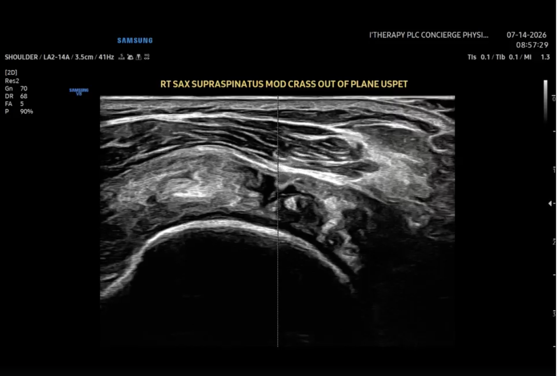
Precision in practice: Rotator Cuff
Recently, our team utilized an out-of-plane, lateral to medial approach for an MSK ultrasound guided percutaneous electrolysis of the supraspinatus. By placing the patient in a supine modified Crass position, we were able to optimize tendon visualization and ensure exact treatment delivery to the tear while keeping the patient comfortable.
We are incredibly grateful to serve the Grand Rapids community. Our clinical philosophy is simple: we believe that prioritizing diagnostic accuracy and highly targeted treatments in every single session ultimately leads to better, more reliable outcomes.
We don't believe in guessing when we can see. If you value a precision based approach to orthopedic care and recovery, our clinic is here to help you get back to doing what you love.
Clinical notes originally published on Dr. Lyons' professional network. [View original case notes.]
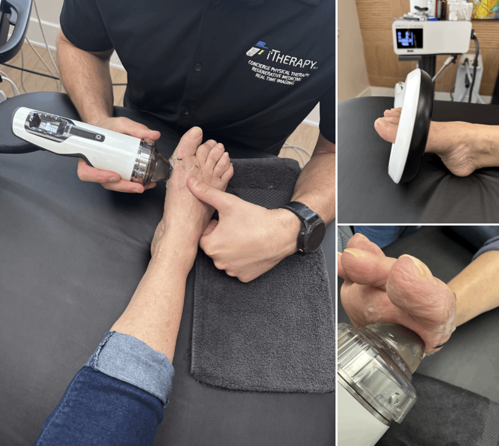
Accelerating recovery when it matters most.
This patient is receiving a combination of fESWT (Focused Shockwave) and EMTT to stimulate osseous integration around surgical hardware/bone and accelerate healing after a 1st MTP joint fusion. Because this patient has osteopenia, proactively mitigating post op complications is our top priority.
What you don’t see in this shot? Before treatment, we use real-time MSK ultrasound to meticulously map out the joint line, screws, and plates. This allows us to direct the regenerative energy exactly where it’s needed most.
The takeaway: We are a niche practice for patients who truly value their time and outcomes. By bridging precision based advanced scope physiotherapy, real-time imaging, and regenerative medicine, we don't just hope for a good outcome.....we engineer it.
Clinical notes originally published on Dr. Lyons' professional network. [View original case notes.]
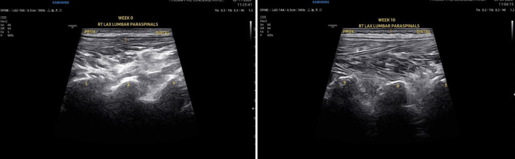
Can we actually REMODEL spinal muscle tissue after chronic fatty infiltration? Yes!
The Case: A complex lumbar patient presenting with gross stability/mobility deficits, SI joint dysfunction, and severe daily pain (VAS 8-9/10). Imaging (US & MRI): Severe bilateral L3-L5 multifidi/rotatores fatty infiltration (hyperechoic signals), L2-3 Modic type 1 changes, SI joint arthritis with spurring, and multi-level disc loss/bulges (L2-L5).
The Protocol: A targeted 10-week program. To ensure ultimate precision, we manually marked the exact spinal segments prior to every single session before applying:
-Focused Shockwave Therapy
-EMTT (Extracorporeal Magnetotransduction Therapy)
-Radial Pressure Wave Therapy
While we are highlighting these energy-based treatments for their role in tissue regeneration, active rehabilitation was equally critical. The patient was carefully progressed in total body mobility and stability exercises week by week, along with the incorporation of a progressive walking program.
The Results:
Objective (See LAX Ultrasound Pics): Went from significant hyperechoic fatty infiltration at Week 0 to COMPLETE remodeling of the multifidi and rotatores by Week 10, restoring normal paraspinal echogenicity!
Subjective & Functional: VAS pain scores plummeted from 8-9/10 down to consistently 1-2/10. The patient noted a 75% improvement in overall pain tolerance across all categories, and is currently operating with ZERO functional limitations.
Clinicians: How are you addressing multifidus atrophy in your complex lumbar patients?
Patients: Structural healing and tissue remodeling are absolutely possible, even with complex MRI findings. Don't lose hope!
Clinical notes originally published on Dr. Lyons' professional network. [View original case notes.]
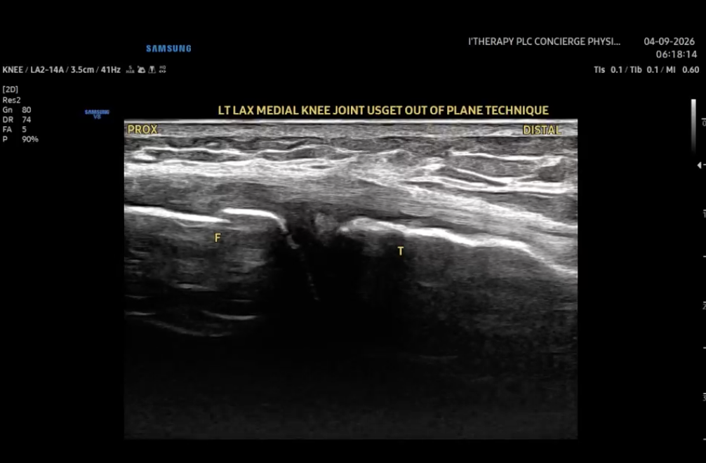
Clinical Case: Out-of-Plane USPET for a Horizontal Medial Meniscus Tear
In the video below, we are demonstrating an out-of-plane approach to Ultrasound-Guided Percutaneous Electrolysis Therapy (USPET) for a horizontal medial meniscus tear (Confirmed on MRI along with associated bone marrow edema). This was a local physician whom wanted to try something different than the well established PRP/BMAC approaches that would allow him less activity restrictions and greater patient comfort during/after the procedure.
Will keep you updated on patient progress.
If you watch closely, you can clearly see the cross-section of the needle "bounce" during fenestration. We use this deliberate technique for enhanced real-time visualization, ensuring the 0.3 x 60mm needle is exactly where it needs to be. As always, the procedure is performed in full sterile fashion.
What about patient comfort? This is where the protocol really shines. Procedure pain is a major barrier for many patients exploring minimally invasive interventions. To combat this, we utilize hyperstimulation radial pressure wave therapy (STORZ RPW Device) directly over the needle entry site prior to the procedure.
The clinical result? Exceptional patient tolerance. Patients consistently report a VAS pain score of 2/10 or less throughout the entire process, including the initial needle entry.
Minimally invasive, highly targeted, and optimized for patient comfort.
I'd love to hear from my network:
Clinicians: Have you experimented with radial pressure wave therapy for pre-procedure analgesia in your US-guided interventions?
Patients: Does knowing a procedure is optimized to stay under a 2/10 pain level change how you view advanced rehab options?
Let me know your thoughts in the comments!
Static shot in the comments sections better demonstrating the horizontal tear and incidental osteophyte and the medial joint line.
Clinical notes originally published on Dr. Lyons' professional network. [View original case notes.]
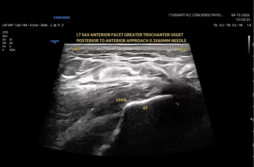
Conquering Lateral Hip Pain: 90% Resolution in Just 6 Weeks
Stubborn lateral hip pain can be incredibly debilitating, but pairing pinpoint ultrasound diagnostics with an advanced, multi-modal treatment plan changes the game.
We recently treated a patient suffering from severe gluteal tendinopathy. Thanks to a highly targeted approach, they achieved a 90% resolution in symptoms and return to function by week 6!
Here is a breakdown of the case and the protocol we used:
The Diagnosis:
Diagnostic MSK Ultrasound revealed a partial-thickness tear of the gluteus minimus tendon right on the anterior facet of the greater trochanter.
The Protocol (Over 6 Weeks):
-Regenerative Intervention: Two Ultrasound-Guided Percutaneous Electrolysis (USGET) sessions, spaced 15 days apart.
-Stacked Energy-Based Therapy: A comprehensive 6-week course utilizing FESWT (Focused Shockwave), RPW (Radial Pressure Wave), and EMTT (Extracorporeal Magnetotransduction Therapy).
-Precision Tracking: MSK ultrasound guidance was used during every visit to map the exact location of the anterior facet and track tendon pathology healing.
For the Clinicians: Technique & Setup
Pre-Treatment Prep: Prior to the USGET, we completed a hyper-stimulation protocol over the anterior facet needle entry point using RPW (21Hz, 2000 shocks, 2.0 bar with a D20T tip) to maximize patient comfort.
The Approach (See Video): MSK ultrasound-guided percutaneous electrolysis using an in-plane approach. We used a 0.3x60mm needle, moving posterior-to-anterior in the short axis directly over the anterior facet of the greater trochanter.
The Patient Experience:
Advanced treatments shouldn't mean extreme pain. Thanks to the hyper-stimulation prep and precision US guidance, the patient’s VAS (pain) scores during the electrolysis procedure maxed out at only 4/10, with an average score of just 2/10!
Clinical notes originally published on Dr. Lyons' professional network.[View original case notes.]
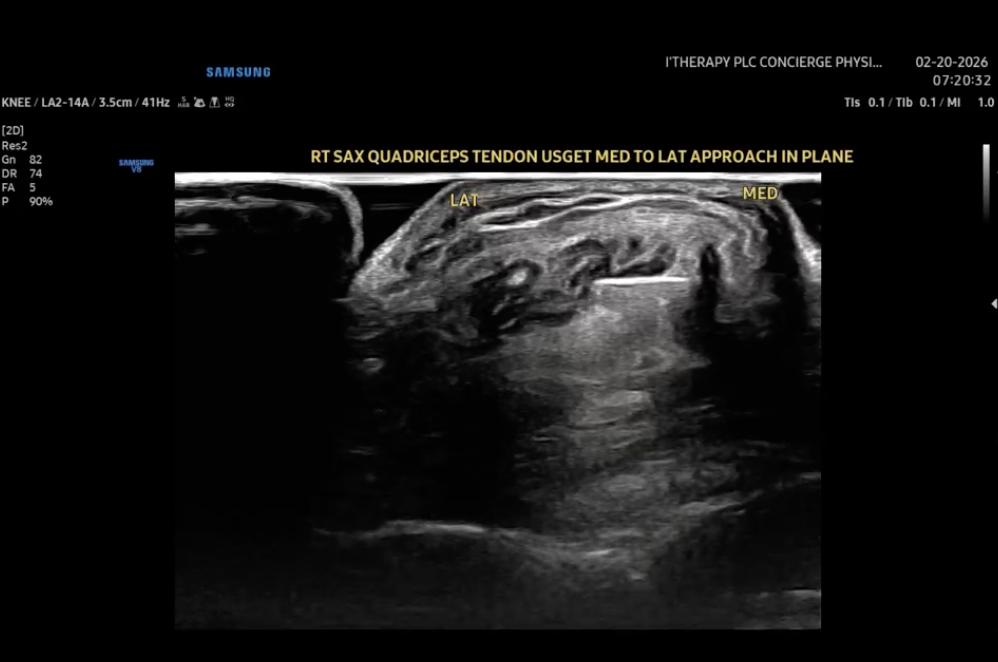
Managing Intrasubstance Tendon Tears
We recently managed a challenging case of Quadriceps Tendinopathy involving a small intrasubstance tear. Tendons are notorious for their slow healing rates, often requiring a push to restart the remodeling process.
Here is a look at the dual-modality approach we used to optimize patient comfort and procedural accuracy:
Phase 1: The Setup (Neuromodulation) Before any invasive work, we utilized Radial Pressure Wave Therapy (RPWT) to induce hyperstimulation analgesia.
-The Protocol: 4000 shocks at 21Hz.
-The Goal: By overwhelming the sensory input (Gate Control Theory), we kept the patient’s VAS (pain score) at a manageable 3/10 or less. This prepared the tissue and the nervous system for the next step.
Phase 2: The Procedure (Precision Electrolysis) With the area desensitized, we moved to ultrasound-guided percutaneous electrolysis.
-Visualization: We used a Short Axis (SAX) in-plane approach, moving medial to lateral.
-Targeting: Using a 0.3mm x 60mm needle, we visualized the tip directly entering the intrasubstance tear.
-Mechanism: The application of a galvanic current targets the degenerated tissue directly, aiming to trigger an acute inflammatory response necessary for regeneration.
It is fascinating to see how combining modalities....using shockwave for pain gating and electrolysis for tissue targeting....can change the game for chronic tendon issues.
Thoughts for the clinicians out there: Do you prefer SAX or LAX (Long Axis) views for your in-plane approaches on the quad? Let’s discuss in the comments.
Clinical notes originally published on Dr. Lyons' professional network.[View original case notes.]
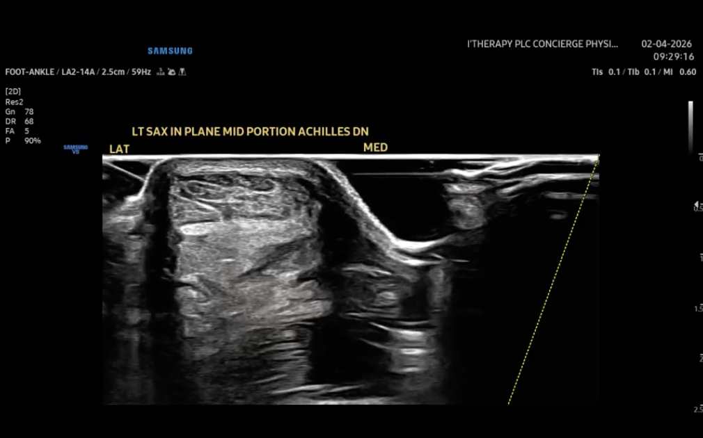
Achilles Tendon Tear: Precision imaging meets comprehensive recovery
Here's a recent case that highlights the value of combining precision guidance with a multimodal, evidence-based approach to tendinopathy.
In the video below, we demonstrate an in-plane, ultrasound-guided dry needling procedure for a partial Achilles tendon tear, clearly visualized using MSK short axis (SAX) imaging from lateral to medial trajectory. Notice needle orientation is set in the small gel stand off to left of video prior to insertion avoiding having to redirect needle trajectory mid procedure (making procedure more comfortable to the patient).
Because tendon healing requires comprehensive support, we didn't stop at the needle. We integrated a stacked protocol of regenerative modalities to optimize tissue repair:
-FESWT (Focused Extracorporeal Shockwave Therapy)
-EPAT (Radial Pulse Therapy)
-EMTT (Extracorporeal Magnetotransduction Therapy)
-Photobiomodulation (Laser Therapy)
The Results: At the patient’s 2-week follow-up, a re-scan via MSK ultrasound showed that the tear had successfully healed. Even better, the patient reported a 50% reduction in their day-to-day symptoms. Patient will now begin eccentric loading phases to increase tensile forces and strengthen collagen deposition/organization.
We are incredibly grateful to play a role in this patient's recovery journey and are always humbled by the body's ability to heal when given the right targeted support.
Always happy to discuss clinical mechanics with colleagues or treatment paths with those looking to finally resolve stubborn tendon injuries. Let’s get you back to doing what you love.
Clinical notes originally published on Dr. Lyons' professional network.[View original case notes.]
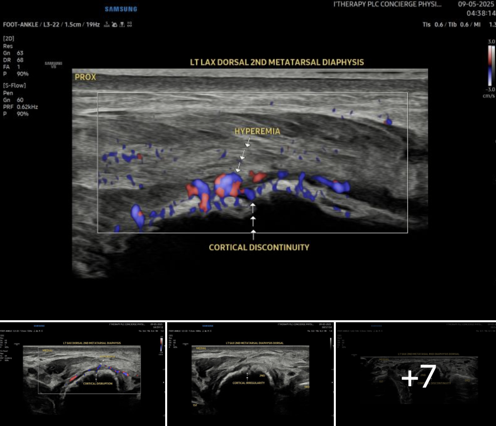
24-Day Complete Union of a 2nd Metatarsal Fracture in a Young Athlete Guided by Real Time Imaging + High-Energy FESWT:
A young, healthy male athlete presented with a 2nd metatarsal fracture and was placed WBAT in a CAM boot on day one.
The included images show the progression from initial cortical irregularities, to cortical closure, and ultimately robust callus formation around the 2nd metatarsal fracture site on X-ray.
Using a precision-based approach, we performed ultrasound imaging in both long and short axis planes prior to every treatment session. This allowed us to target high-energy FESWT (0.35 mJ/mm²) directly at the fracture line with accuracy and consistency each visit.
After 24 days, the patient demonstrated:
-Complete unionization of the fracture
-Robust circumferential callus formation
-Pain free return to progressive loading
Treatment Frequency: 2x/week
Protocol Per Visit:
-FESWT: 6,000 shocks @ 0.35 mJ/mm²
-EMTT: 20,000 shocks | Level 8 | 8 Hz
-PEMF (Thoracic) + Red/Infrared Photobiomodulation Panel
-PBFR: Progressive load tolerance, pain-free, 5 groupings @ 80% LOP
This case reinforces the impact of combining real-time imaging, mechanotransductive therapies, and intelligent loading strategies to accelerate recovery in high-demand individuals.
Clinical notes originally published on Dr. Lyons' professional network.[View original case notes.]
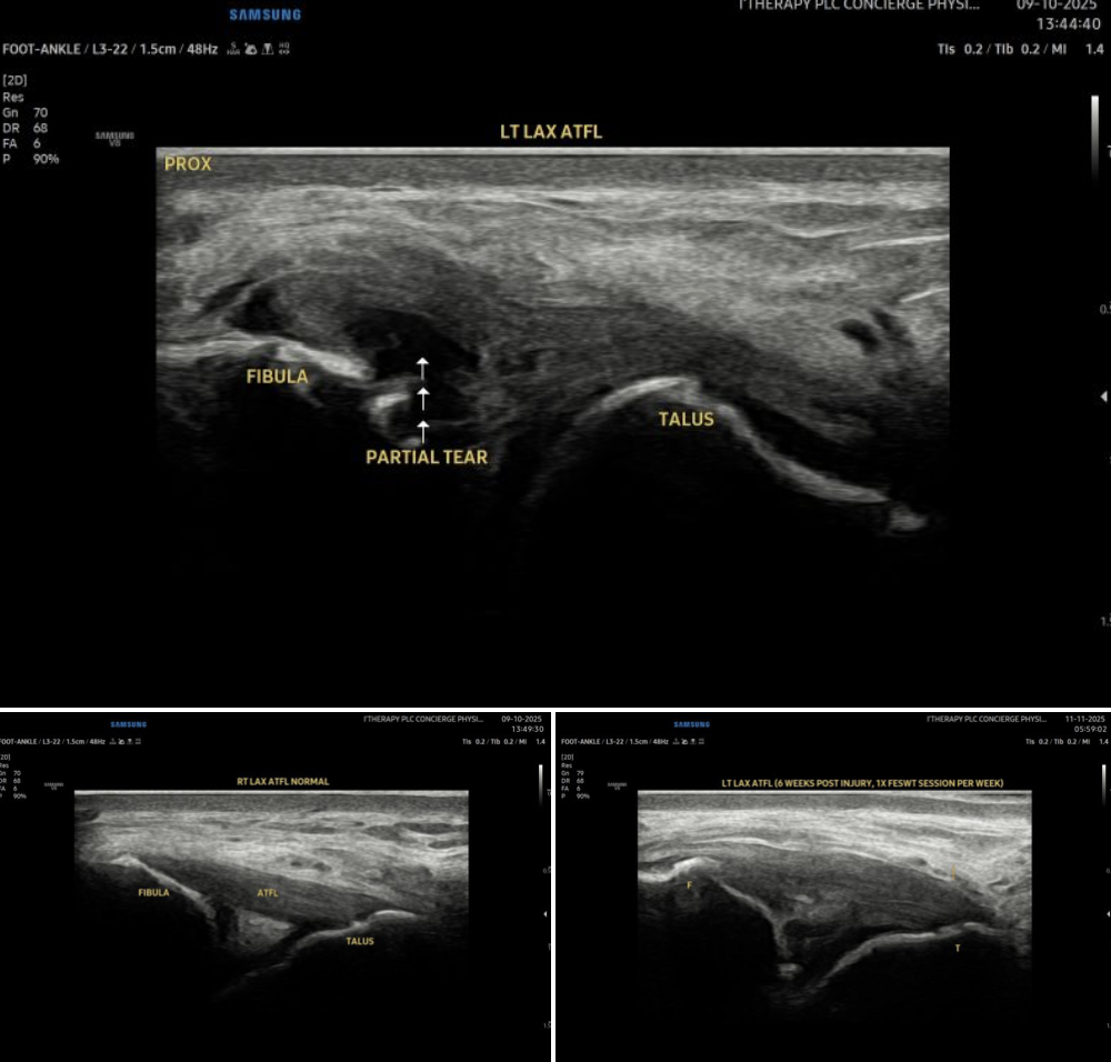
Ultrasound-Proven Ligament Healing After 6 Weeks of F-ESWT and EMTT Combination Therapy
6 weeks ago, a 14-year-old football player presented after a hard ankle inversion injury..... ultrasound revealed a partial tear of the anterior talofibular ligament (ATFL).
Our treatment approach combined:
-Focused Extracorporeal Shockwave Therapy (F-ESWT): 1x/week × 6 weeks (3,000 shocks; 0.15–0.25 mJ/mm²)
-Electromagnetic Transduction Therapy (EMTT): 10,000 pulses at level 8, 8 Hz
At week 6, ultrasound confirmed healing of the ATFL, with full return to pain-free function and sport participation. You will notice that although healed the ligament is still thickened.
While each case is unique, this highlights how noninvasive regenerative modalities.....when applied with precision and guided by imaging....can accelerate soft tissue recovery and reduce downtime in young athletes.
(Images: Week 0 vs. Week 6.....partial ATFL tear to full resolution, with normal non-involved side for baseline comparison).
Clinical notes originally published on Dr. Lyons' professional network.[View original case notes.]